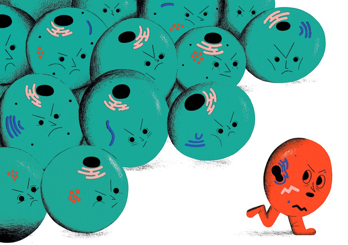
Yasuyaki Fujita has seen first-hand what happens when cells stop being polite and start getting real. He caught a glimpse of this harsh microscopic world when he switched on a cancer-causing gene called Ras in a few kidney cells in a dish. He expected to see the cancerous cells expanding and forming the beginnings of tumours among their neighbours. Instead, the neat, orderly neighbours armed themselves with filament proteins and started “poking, poking, poking”, says Fujita, a cancer biologist at Hokkaido University in Sapporo, Japan. “The transformed cells were eliminated from the society of normal cells,” he says, literally pushed out by the cells next door.
In the past two decades, an explosion of similar discoveries has revealed squabbles, fights and all-out wars playing out on the cellular level. Known as cell competition, it works a bit like natural selection between species, in that fitter cells win out over their less-fit neighbours. The phenomenon can act as quality control during an organism’s development, as a defence against precancerous cells and as a key part of maintaining organs such as the skin, intestine and heart. Cells use a variety of ways to eliminate their rivals, from kicking them out of a tissue to inducing cell suicide or even engulfing them and cannibalizing their components. The observations reveal that the development and maintenance of tissues are much more chaotic processes than previously thought. “This is a radical departure from development as a preprogrammed set of rules that run like clockwork,” says Thomas Zwaka, a stem-cell biologist at the Icahn School of Medicine at Mount Sinai in New York City.
But questions abound as to how individual cells recognize and act on weaknesses in their neighbours. Labs have been diligently hunting for — and squabbling over — the potential markers for fitness and how they trigger competitive behaviours. These mechanisms could allow scientists to rein in the process or to help it along, which might lead to better methods for fighting cancer and combating disease and ageing using regenerative medicine.
“Cell competition is on the global scientific map,” says Eugenia Piddini, a cell biologist at the University of Bristol, UK, who likens the buzz around this idea to the excitement that helped propel modern cancer immunotherapies. The better scientists understand competition, she says, the more likely it is that they will be able to use it therapeutically.
History repeats
During a blizzard that dumped more than 30 centimetres of snow this past February, biologists from about a dozen disciplines convened at a hotel at Lake Tahoe, California, for the first major meeting devoted to cell competition.
“It was a zoo of researchers,” says co-organizer Zwaka, and included biologists who study flatworms that can regenerate their whole body from a single cell, geneticists attempting to make interspecies chimaeras of mouse, monkey and rabbit embryos, and a keynote speaker who spoke about the terrible battles and cooperative campaigns waged in bacterial communities.
The snowbound attendees, about 150 in all, debated how and why cells size up their competition. And they celebrated the discovery that gave birth to the field.
How secret conversations inside cells are transforming biology
In 1973, two PhD students, Ginés Morata and Pedro Ripoll were perfecting a way to track the various cell populations in a fruit-fly larva that would eventually develop into a wing. Working at the Spanish National Research Council’s Biological Research Center in Madrid, they introduced a mutation called Minute into a few select cells in the larva and left the rest of the cells unaltered.
Knowing that Minute cells grow slower than their unaltered neighbours, the scientists expected to find some smaller cells amid the wild-type counterparts. “Instead, we found that the cells disappeared,” says Morata, now a developmental biologist at the Autonomous University of Madrid in Spain.
On their own, Minute cells can develop into a fly that is normal — except for the short, thin bristles on their bodies that give the mutation its name. But when mixed with wild-type cells in the larva, the cells simply vanished. “Minute cells were not able to compete with the more vigorous, metabolically active wild-type cells,” says Morata. They described the activity as cell competition. “It was a very surprising and interesting observation,” Morata says. But lacking the molecular tools to follow cell fates more closely, he and his colleagues let the finding simmer.
Twenty-six years later, postdocs Laura Johnston and Peter Gallant observed nearly the same phenomenon. Working with Bruce Edgar and Robert Eisenman, respectively, at the Fred Hutchinson Cancer Center in Seattle, Washington, they were studying a mutation in another fly gene, Drosophila Myc (dMyc), that also slows cell growth.
“There was a eureka moment when Peter and I realized that these dMyc mutant cells would disappear,” says Johnston, now a developmental biologist at Columbia University Medical Center in New York City. They eventually showed that the mutant cells were forced to initiate a form of programmed cell death called apoptosis. “It was very clear that this was a competitive situation,” Johnston says.
Their 1999 paper ignited interest among scientists, including Morata. He jumped back into the fray with Eduardo Moreno, and they took advantage of modern molecular tools to repeat the Minute experiments. “The field blossomed from there,” says Johnston.
Myc acts as a master controller of cell growth, and Minute encodes a key component needed for synthesizing proteins — so it’s not surprising that reduced expression of those proteins makes cells less fit. But Johnston’s next finding took people by surprise. She showed that cells with an extra copy of normal dMyc outcompeted wild-type cells. These fitter-than-wild-type cells came to be called “supercompetitors”.
Johnston’s discovery of supercompetition emphasized that cell competition is about the relative fitness of a group of cells, says Zwaka. If one cell is falling behind, the entire group of neighbours could decide it has to go. But on the flipside, they can also sense that certain cells are better and should survive.
Cell competition wasn’t simply about getting rid of defects; it was about survival of the fittest, with the less-fit ‘loser’ cells dying and the ‘winners’ proliferating. Importantly, competition was seen only when there was a mixture of genetically different cells, a phenomenon known as mosaicism. In this way, cell competition acts like a quality-control system, booting out undesirable cells during development.
Vying for viability
Fujita’s observation of the kicked-out kidney cells was one of the first hints that mammalian cells compete, too. Soon after that work was published, researchers started to observe competition forcing out mutated cells from various other tissue types such as skin, muscle and gut.
The next most obvious place to look for competing cells was the mammalian embryo. In 2013, Zwaka’s team, and two other laboratories, probed mouse embryos at the earliest stage of development — those that have progressed just beyond a ball of cells. Zwaka’s group made mouse embryonic stem cells (ESCs) with a supercompetitor mutation that lowered expression of p53, an important quality-control protein that normally puts the brakes on cell division. When these cells were put into a mouse embryo, they quickly took over and developed into a normal mouse5. Similarly, Miguel Torres’s lab at the National Center for Cardiovascular Research in Madrid showed that supercompetition could be induced in an early mouse embryo using slight overexpression of the mouse Myc gene.
By artificially creating losers or winners, researchers could force cell competition into play. But Torres’s team, led by then-postdoc Cristina Clavería, also made the striking observation that Myc expression varied naturally in mouse ESCs. Cells in the embryo with approximately half the amount of the protein compared with their neighbours were dying by apoptosis. This was one of the first studies that strongly pointed to naturally arising cell competition.
Sculpting tissues
The phenomenon also comes into play later on in embryonic development. In a study published this year, postdoc Stephanie Ellis at Elaine Fuch’s lab in Rockefeller University in New York City, looked at mouse skin. During development, its surface area expands by a factor of 30 over the course of about a week. The cells within proliferate wildly — first as a single layer and later as multiple layers.
Ellis injected mouse embryos with a concoction that turns cells into genetic losers. She targeted a few cells present when the embryonic skin is a single layer thick, and added a marker gene that made them glow red. Then she used time-lapse imaging to watch their grim fates: the skin cells popped out from the surface layer, broke up and disappeared. Later, she noticed the winner cells engulfing and clearing the losers’ corpses.
Repeating the experiment at the multilayer stage, Ellis no longer saw the less-fit skin cells perishing or being engulfed. Instead, the loser cells tended to differentiate and migrate into the outer layers of skin — where they acted as a barrier for a short time before being shed. The winner cells were more likely to remain behind in the bottom layer as stem cells.
This made sense. “Killing a cell is energetically expensive,” says Ellis. A developing tissue, she says, might decide: “Why not just remove losers through differentiation?” Emi Nishimura’s lab at the Tokyo Medical and Dental University in Japan, found that competing stem cells in the ageing tail skin of adult mice used the same pattern of asymmetrical divisions to eliminate stem cells with lower levels of a key structural collagen protein
This made sense. “Killing a cell is energetically expensive,” says Ellis. A developing tissue, she says, might decide: “Why not just remove losers through differentiation?” Emi Nishimura’s lab at the Tokyo Medical and Dental University in Japan, found that competing stem cells in the ageing tail skin of adult mice used the same pattern of asymmetrical divisions to eliminate stem cells with lower levels of a key structural collagen protein.
These experiments could provide guidance for scientists looking to harness stem cells to rejuvenate ageing tissues and organs. Cell competition could either help or hurt such therapies: stem cells might outcompete older, less-fit cells, or they might encounter a hostile neighbourhood when transplanted into tissue. Understanding whether and how cell competition happens in adult tissue could help settle this matter.
Piddini admits that she was a little obsessed with the idea, and her group was part of a wave of researchers that proved cell competition does take place in adult organisms. To test the idea, she says, the team “genetically sprinkled” a mutated copy of RPS3, a gene functionally related to Minute, into some cells in the intestine of adult flies. Cells with the mutant copy were outcompeted by their wild-type counterparts. It didn’t matter whether the losers were the stem cells that maintain the gut or differentiated cells: all eventually perished.
Cristina Villa del Campo, a senior postdoc in the Torres lab, tested for adult competition in the mouse heart by introducing winner cardiac cells at eight to ten weeks of age. Over the course of one year, she tracked the numbers of winner cells and wild-type losers and saw the loser population decline by about 40%.
“It was a slow replacement in the adult,” says Villa del Campo. “But even highly differentiated functional adult cells can sense the less-fit heart cells and eliminate them.”
Unanswered questions
Even with so many examples of cell competition playing out in different conditions, the field still faces a torrent of unanswered questions. One big puzzle is how cells in a group sense fitness. “Maybe cells are recognizing chemical differences, or physical differences, or differences in cell-membrane composition,” says Fujita, who adds that labs have found evidence for all three.
His filament-poking kidney-cell experiments suggest that cell–cell contact is needed. Others have seen chemical-fitness signals that seem to be short-range, travelling up to eight cell diameters. Exactly which molecules are responsible for this signalling — either secreted chemicals or physical tags — is the subject of intense debate and investigation.
The secret social lives of viruses
Both Johnston and Zwaka have turned up signals associated with immune surveillance. Johnston’s group identified molecules that typically call immune cells to swarm in and engulf foreign invaders and that were driving death in losers11. Normal cells express low levels of these death signals at all times. But in a competitive mix, winners flooded their loser neighbours with the signal, which pushed them to kill themselves.
Zwaka proposes that cells might assess each other’s health by sniffing out the general signals or debris that cells shed. It’s akin to smelling the steaks that your neighbour is grilling for dinner and concluding that they must be doing well.
Or it could be as simple as seeing which flag your neighbour is flying. Moreno heads his own group now at the Champalimaud Centre for the Unknown in Lisbon, Portugal, which discovered a membrane-spanning protein called Flower. In humans, the protein can take four forms, each displaying its own characteristic structure on the outer cell surface. Two signal ‘I’m a winner’ and the other two signal ‘I’m a loser’ to nearby cells, says Moreno.
Some human cancer cells fly the Flower-winner signals, which might enhance their survival. Experiments in Moreno’s lab showed that silencing the winning flags on tumours slowed the cells’ growth and made them susceptible to chemotherapy.
Some researchers, however, dispute the importance of the Flower tags. Moreno acknowledges that they are not present in all cell-competition situations.
Healthy competition
Cracking the mechanics of competition will be key if researchers want to use it to improve cell-based cancer or regenerative therapies.
There are tantalizing hints that cell competition might already protect against cancer. Findings made in the past few years reveal that human skin, oesophageal and lung cells show high levels of mosaicism. Approximately one-quarter of skin cells, for example, harbour many precancerous mutations that only rarely turn into tumours.
It is unclear what gives cancerous cells the advantage when tumours do form. If researchers can learn how to subdue supercompetitors or blunt cancer cells’ ability to compete, they might be able to turn that against cancer.
Conversely, stem cells might need to gain a competitive edge if they are to replace aged or diseased tissue for an organ makeover. Villa del Campo says that clinicians are already considering how to enhance patient-derived cardiac stem cells to efficiently replace cells that have been damaged by heart attacks or disease.
What started as modest observations in minuscule fruit-fly larvae has exposed the primal cellular battles that could usher in a new era of cell-based medicine. The process has scientists buzzing, but it remains mysterious.
“Cell competition might be a general process to remove any undesirable cell that should not be there,” says Morata, after returning from a one-day meeting in Lausanne, Switzerland devoted to competition in September.
Now , he’s thrilled that work he essentially shelved more than 40 years ago is gaining new life and that the competition is heating up. “It’s really exciting.”
Nature 574, 310-312 (2019)